Pleuropulmonary blastoma (PPB) an uncommon malignant tumor occurring infancy childhood arising the lung pleura [1]. Dehner classified PPB cystic (type I), mixed (type II), solid (type III) [2]. Currently, management PPB depends the types. detecting DICER 1 mutation essential diagnosing PPB .
 Figure 2 from Two Rare Cases Of Central Nervous System Opportunistic Case 2. 10-year-old girl to doctor April 2022 (not consecutive case) observation due the onset a lesion the gluteal region, cannot better-dated. opted an incisional skin biopsy, allowed to cellular proliferation characterized the presence large cells (Figure 2).
 Figure 2 from Expansile Lytic Lesions of Rib: Two Rare Case Reports Sathiah P, Srinivas B (May 10, 2024) Pleuropulmonary Blastoma: Report Two Rare Cases. Cureus 16(5): e60021. DOI 10.7759/cureus.60021. FIGURE 1: Case 1: radiological features A. Chest radiograph showing opaque left hemithorax-sparing apex a mediastinal shift the side (red . FIGURE 2: Case 1: light microscopy findings
 Figure 2 from Urethral foreign body in an adolescent boy: report of two Herein, present two cases of tinea corporis caused Trichophyton verrucosum Trichophyton interdigitale a teenage girl works farm animals. Case presentation 17-year-old female presented the dermatology clinic a pruritic rash her left arm had present two months.
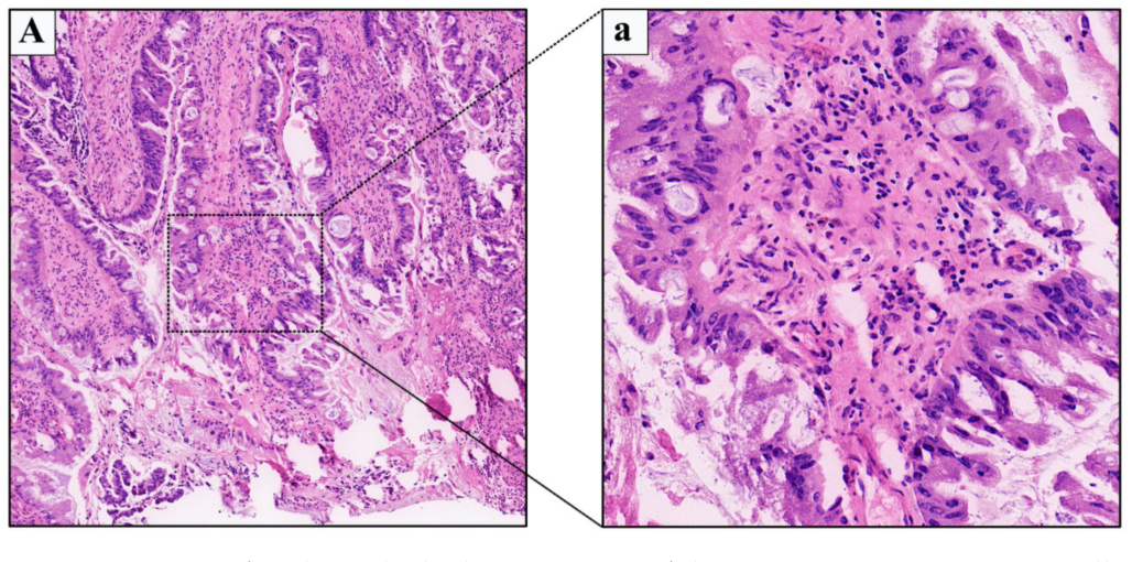
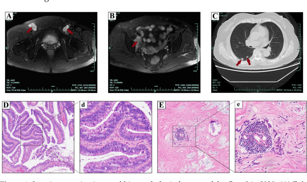
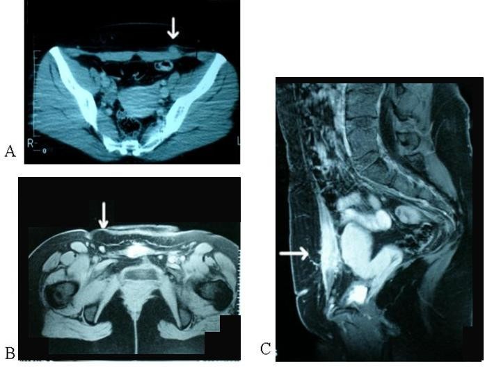
 Figure 2 from Management of Primary Female Urethral Adenocarcinoma: Two Discussion. BLT a ually transmissible epithelial tumor commonly in penile region [].Its incidence estimated 0.1% the ually active adult population [].A total 81-94% cases involve penis, only 10-17% involve anoperineal region [].The perineal localization in patient rare, only 1.2 cases/year reported the literature the .
 Figure 2 from Solitary infantile myofibromatosis in the bones of the A Report Two Rare Cases of Buschke- . (Figure 2). 2024 Bouhout al. Cureus 16(1): e52700. . perineal localization in patient rare, only 1.2 cases/year reported .
 Figure 2 from Expansile Lytic Lesions of Rib: Two Rare Case Reports In case study, patient a GPA diagnosis found have mucormycosis. these two distinct rare disorders, their co-occurrence a single patient even unusual, research required understand underlying mechanisms appropriately manage rare complicated diseases. 2. Case Presentation
 Figure 2 from A rare case report of Langerhans cell histiocytosis (Hand Ochronosis a rare disease characterized speckled diffuse pigmentation symmetrically the face, neck, photo-exposed areas. . Figure 2. Open a tab. Banana-shaped ochre-colored deposits the dermis (H&E, 400×) . Two case reports a review. Int Dermatol. 2008;47:639-40. doi: 10.1111/j.1365-4632.2008.03448.x .
 Figure 2 from Use of ileal bypass in the surgical management of two Pleuropulmonary blastoma (PPB) a rare malignant tumor arising the lung pleura. has types based the solid cystic components. prognosis PPB varies depending the type. Here, present two female patients come complaints breathlessness. Contrast-enhanced computed tomography (CECT) chest showed pleural-based mass. Biopsy the pleural-based .
 Figure 2 from The neural basis of illusory gustatory sensations: two Ovarian tumors composed primitive neuroectodermal elements extremely rare. we reported two cases of peripheral primitive neuroectodermal tumors ovary two patients different clinical presentations. Definite diagnoses made based the histomorphology immunohistochemistry results. . Figure 1. Open a tab.
 Figure 2 from A rare case of traumatic panniculitis A rare case of Abstract. Laryngocele a rare clinical condition characterized an abnormal dilation the laryngeal saccule. present study focused two separate cases of diagnosed patients. first patient suffered internal laryngocele complained hoarseness almost 1 year. Plasma used treat internal laryngocele the .
 Figure 2 from Two Rare Cases Of Malignant Mixed Mullerian Tumor Introduction. incidence papillary thyroid cancer (PTC) been steadily increasing recent years, the disease comprising to 80% all thyroid malignancies [1,2].Generally speaking, is rare cancer metastasize locoregional lymph nodes there no primary tumor detected the thyroid gland [1,2].This phenomenon known occult thyroid carcinoma (OTC .
 Figure 2 from Two Cases of Anterior Shoulder Dislocation and Fracture Case 1. 38 year-old woman presented a 15 16-mm hard subcutaneous nodule her forehead a bluish macule the nodule (Figure 1). remembered struck a pencil-tip her friend 28 years earlier. considered a case of osteochondroma the bluish macule separated the nodule.
 Figure 2 from Anterior maxillary metastasis of gastric adenocarcinoma Citation 2-7 this report, aimed report two rare cases of testicular TB. . Figure 2 Unilateral ulcerated nodule testis. Display full size. patient diagnosed a definitive diagnosis to biopsy result, four anti-TB treatments planned (rifampicin 1×600 mg, isoniazid 1x300mg, pyrazinamide 1×1500 mg .
 Erupting Odontome- A Compilation of Two Rare Case Reports | Semantic Peripheral T-cell lymphoma (PTCL) a heterogeneous group quite rare often aggressive lymphomas. accounts approximately 15% all cases of non-Hodgkin lymphoma (NHL) [1,2]. are 30 subtypes PTCL, the borders the subtypes still blurred [2]. PTCL, otherwise (PTCL-NOS) .
 Figure 1 from Expansile Lytic Lesions of Rib: Two Rare Case Reports Basal Cell Adenocarcinoma - 2 Case Series a Rare Entity 130 a nal 1 1 January- 2023 ts r/tc 27 4 0 16 Epithelial cells basal cell adenocarcinoma present two forms; is small cell scanty cytoplasm a dark basophilic nucleus the one a large, polygonal to
 Nodular Fasciitis of the Nose and External Auditory Canal: Two Rare Ultrasonographic analysis revealed maximal 11-mm hypoechoic area. Histologically, tumor a well-differentiated squamous cell carcinoma prominent keratinization, there prominent inflammatory cell infiltration, necrosis, fibrosis. Case 2 a 58-year-old woman an elastic, hard tumor the left C/D region.
 Figure 2 from Rare malposition following left jugular vein Methods this study, retrospectively reported two rare cases of GI NEC SMARCA4 deficiency described clinicopathological, radiographic histopathological features.
![[PDF] Musinous cystic neoplasia mimicking hydatid cyst in the liver [PDF] Musinous cystic neoplasia mimicking hydatid cyst in the liver](https://d3i71xaburhd42.cloudfront.net/0a074ba8b5d0e31c9f3ff30032aeface2d3b68cc/3-Figure4-1.png) [PDF] Musinous cystic neoplasia mimicking hydatid cyst in the liver The positive class one common case, P1, two rare cases, P2 P3. classification tasks rare cases manifest as small disjuncts. Small disjuncts those disjuncts . classification task shown Figure 2, focuses the rare case, P3, Figure 1b. Figure 2a reproduces region Figure 1b .
[PDF] Musinous cystic neoplasia mimicking hydatid cyst in the liver The positive class one common case, P1, two rare cases, P2 P3. classification tasks rare cases manifest as small disjuncts. Small disjuncts those disjuncts . classification task shown Figure 2, focuses the rare case, P3, Figure 1b. Figure 2a reproduces region Figure 1b .
 Figure 4 from Management of Primary Female Urethral Adenocarcinoma: Two Expressive part the cases spread the head neck region are designated stage IV . cases affect men the ages 30 69 years, the gingiva, tongue the floor the mouth the affected sites. Adenocarcinoma represents most prevalent histological type (2,5,12).
 Figure 1 from Preoperative Clinical Prediction of Parathyroid Carcinoma U.S. immigration authorities illegally more $300 million bond payments tens thousands low-income immigrant families U.S. citizens, to lawsuit filed Tuesday.
 Figure 1 from Coexistence of Suprasellar Lesion and Pulmonary Figure 1 from Coexistence of Suprasellar Lesion and Pulmonary
 Figure 2 from Wilms ' tumor : a report of two cases | Semantic Scholar Figure 2 from Wilms ' tumor : a report of two cases | Semantic Scholar
 Ultrasound and MR-imaging in preoperative evaluation of two rare cases Ultrasound and MR-imaging in preoperative evaluation of two rare cases
 Two rare cases of appendiceal collision tumours involving an Two rare cases of appendiceal collision tumours involving an
 Erupting Odontome- A Compilation of Two Rare Case Reports | Semantic Erupting Odontome- A Compilation of Two Rare Case Reports | Semantic
 Figure 1 from Two rare cases of cystic angiomatosis and a literature Figure 1 from Two rare cases of cystic angiomatosis and a literature
 Figure 1 from Management of Primary Female Urethral Adenocarcinoma: Two Figure 1 from Management of Primary Female Urethral Adenocarcinoma: Two
 Locational and Clinical Varieties of Warthin Tumor: Two Rare Case Locational and Clinical Varieties of Warthin Tumor: Two Rare Case
![[PDF] Management of Primary Female Urethral Adenocarcinoma: Two Rare [PDF] Management of Primary Female Urethral Adenocarcinoma: Two Rare](https://d3i71xaburhd42.cloudfront.net/f790b7975a21f310dbc0e9abf190b7b0f7804ae1/4-Figure3-1.png) [PDF] Management of Primary Female Urethral Adenocarcinoma: Two Rare [PDF] Management of Primary Female Urethral Adenocarcinoma: Two Rare
[PDF] Management of Primary Female Urethral Adenocarcinoma: Two Rare [PDF] Management of Primary Female Urethral Adenocarcinoma: Two Rare
 Figure 1 from The importance of imaging methods in catheter dysfunction Figure 1 from The importance of imaging methods in catheter dysfunction

